Bones In Your Back Diagram : Muscles Of The Lumbar Spine Of The Trunk - Spinal anatomy is a remarkable combination of strong bones, flexible ligaments and tendons, large muscles and highly sensitive nerves.
Bones In Your Back Diagram : Muscles Of The Lumbar Spine Of The Trunk - Spinal anatomy is a remarkable combination of strong bones, flexible ligaments and tendons, large muscles and highly sensitive nerves.. This process continues until the end of puberty, when the growth plate stops growing and the bones fuse permanently into a single bone. In the back and elsewhere in the body, tendons attach muscles to bones. It contains the osteology, arthrology and myology of the spine and back. The knee joint is the largest joint in the body and is primarily a hinge joint, although some sliding and rotation occur. The pubis, ischium, and ilium together constitute the pelvis while the thigh bone is the femur.
Fpe medical review board a foot pain diagram is a great tool to help you work out what is causing your ankle and foot pain. The spine runs from the base of your skull down the length of your back, going all the way down to your pelvis. Bone spurs do not necessarily lead to lower back pain, but they are a common cause of it. You have 33 vertebrae (bones) that make up the vertebral column. 12 photos of the human back bone chart.
Human Back Bone Chart Back Bones Diagram Human Anatomy Diagram Human Bones Anatomy Human Anatomy Human Back from i.pinimg.com Bone structure birds 12 photos of the bone structure birds bone structure birds, bone structure in. Your spinal cord is protected by the vertebral column (spinal column or backbone). The pubis, ischium, and ilium together constitute the pelvis while the thigh bone is the femur. Tendons connect the knee bones to the leg muscles that move the knee. The muscles of the lower back help stabilize, rotate, flex, and extend the spinal column, which is a bony tower of 24 vertebrae that gives the body structure and houses the spinal cord. There are a whole range of structures e.g. How many bones make up the human spine? It is designed to be incredibly strong, protecting the highly sensitive nerve roots, yet highly flexible, providing for mobility on many different planes.
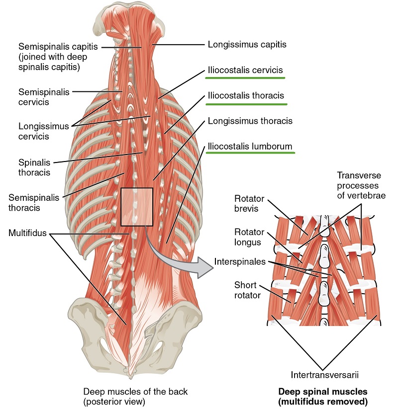
At the back of each bone in the spine (vertebra) are bony points called processes, which muscles attach to.
The bones of the pelvis and lower back work together to support the body's weight, anchor the abdominal and hip muscles, and protect the delicate vital organs of the vertebral and abdominopelvic cavities. At the same time the bones grow larger by growing back into the growth plates. Select your language the spine diagram. It is composed of 300 bones at birth, but later decreases to 80 bones in the axial skeleton and 126 bones in the appendicular skeleton. When most people mention their back, what they are actually referring to is their spine. We are pleased to provide you with the picture named anatomy of back muscles diagram.we hope this picture anatomy of back muscles diagram can help you study and research. Bone diagram forehead (frontal bone) nose bones (nasals) cheek bone (zygoma) upper jaw (maxilla). Bone spurs do not necessarily lead to lower back pain, but they are a common cause of it. Just need a glimpse, leave your valuable advice let us know , and subscribe us! The foot bones shown in this diagram are the talus, navicular, cuneiform, cuboid, metatarsals and calcaneus. Too much joint cracking may damage the joint tissues and accelerate a type of arthritis known as osteoarthritis (the wear and tear type). First, we're going to look at the bone structures. At the back of each bone in the spine (vertebra) are bony points called processes, which muscles attach to.
The spine runs from the base of your skull down the length of your back, going all the way down to your pelvis. Stretch your back muscles first. Bone spurs, or osteophytes, are bony growths that can develop on the spine due to wear and age. Bone diagram forehead (frontal bone) nose bones (nasals) cheek bone (zygoma) upper jaw (maxilla). This human anatomy module is composed of diagrams, illustrations and 3d views of the back, cervical, thoracic and lumbar spinal areas as well as the various vertebrae.
Intrinsic Back Muscles Anatomy Of The Torso Medical Library from d3uigcfkiiww0g.cloudfront.net The five metatarsals are the long bones that link the tarsal bones to the toes, seen in yellow in the diagram below. As you can see from the image below, your back, or spine, is made up of many parts. When most people mention their back, what they are actually referring to is their spine. Bone spurs, or osteophytes, are bony growths that can develop on the spine due to wear and age. They help support particular bones and make them move. Muscle or tendon injuries can occur anywhere in the body. Bone spurs do not necessarily lead to lower back pain, but they are a common cause of it. Fpe medical review board a foot pain diagram is a great tool to help you work out what is causing your ankle and foot pain.
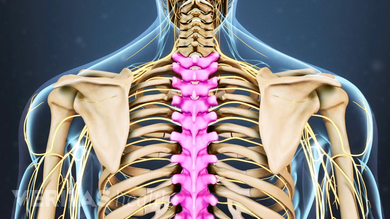
Your spinal cord is protected by the vertebral column (spinal column or backbone).
Spinal anatomy is a remarkable combination of strong bones, flexible ligaments and tendons, large muscles and highly sensitive nerves. In the back and elsewhere in the body, tendons attach muscles to bones. Bones of the pelvis and lower back. The pubis, ischium, and ilium together constitute the pelvis while the thigh bone is the femur. At the back of each bone in the spine (vertebra) are bony points called processes, which muscles attach to. The muscles of the lower back help stabilize, rotate, flex, and extend the spinal column, which is a bony tower of 24 vertebrae that gives the body structure and houses the spinal cord. Spinal fractures can also affect the other parts of the spine—the nerves, spinal cord, ligaments, etc.—and this article will discuss those later. Too much joint cracking may damage the joint tissues and accelerate a type of arthritis known as osteoarthritis (the wear and tear type). The spine is made of 33 individual bones stacked one on top of the other. How many bones make up the human spine? Lumbar spine anatomy diagram images. You have 33 vertebrae (bones) that make up the vertebral column. The foot bones shown in this diagram are the talus, navicular, cuneiform, cuboid, metatarsals and calcaneus.
The muscles, bones, ligaments, and tendons in the back can all be injured and cause back pain. The bones of the leg are the femur, tibia, fibula and patella. The bones of the pelvis and lower back work together to support the body's weight, anchor the abdominal and hip muscles, and protect the delicate vital organs of the vertebral and abdominopelvic cavities. Our latest youtube film is ready to run. There are a whole range of structures e.g.
Spine Anatomy Overview Video from embed.widencdn.net They hold up your body, and. Human backbone diagram, bone, human backbone diagram. As you can see from the image below, your back, or spine, is made up of many parts. The bones of the pelvis and lower back work together to support the body's weight, anchor the abdominal and hip muscles, and protect the delicate vital organs of the vertebral and abdominopelvic cavities. For more anatomy content please follow us and visit our website: This process continues until the end of puberty, when the growth plate stops growing and the bones fuse permanently into a single bone. Select your language the spine diagram. Bone structure birds 12 photos of the bone structure birds bone structure birds, bone structure in.
Bones of the pelvis and lower back.
This spinal column provides the main support for your body, allowing you to stand upright, bend, and twist, while protecting the spinal cord from injury. As you can see from the image below, your back, or spine, is made up of many parts. Bone diagram forehead (frontal bone) nose bones (nasals) cheek bone (zygoma) upper jaw (maxilla). The muscles, bones, ligaments, and tendons in the back can all be injured and cause back pain. Your spinal cord is protected by the vertebral column (spinal column or backbone). Select your language the spine diagram. It contains the osteology, arthrology and myology of the spine and back. The vertebral column of the lower back includes the five lumbar vertebrae, the sacrum, and the coccyx. The five metatarsals are the long bones that link the tarsal bones to the toes, seen in yellow in the diagram below. The first seven bones (vertebrae) of your spine form your neck. First, we're going to look at the bone structures. The spine runs from the base of your skull down the length of your back, going all the way down to your pelvis. Too much joint cracking may damage the joint tissues and accelerate a type of arthritis known as osteoarthritis (the wear and tear type).
The bones together make up the hip back bones diagram. The vertebral column of the lower back includes the five lumbar vertebrae, the sacrum, and the coccyx.


0 Komentar